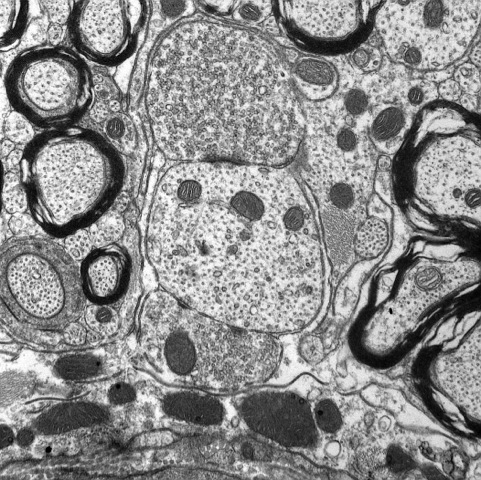
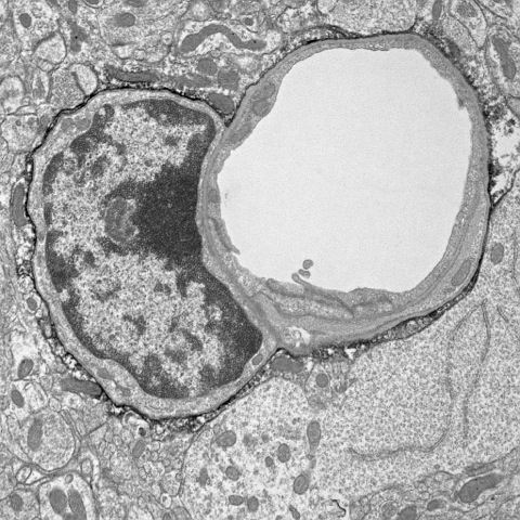
The BMRI is now home to the Neuroanatomy Electron Microscopy (EM) Core. "The Electron Microscopy & Histology Core" provides similar services as the "Neuroanatomy EM Core". The difference is that the Neuroanatomy EM Core focuses on brain and provides training to people (especially students and post-docs) in laboratory procedures. For more information about the entire list of core facilities offered at WCMC, please click here: https://brainandmind.weill.cornell.edu/core-facilities/neuroanatomy-em-core
Summary
The goal of the Core is to provide training and services in the processing of brain tissue for light and electron microscopic immunocytochemistry. Key aspects of the core include: 1) one-on-one training classes; 2) assistance with brain fixation and histology; 3) assistance with quantitative light microscopic immunocytochemistry on brain tissue; 4) assistance with quantitative single and dual labeling electron microscopic immunocytochemistry; 5) assistance with collection, analysis and interpretation of these preparations; and 6) assistance with preparing collected data for publication and meeting presentations.
Mission of the core
This Core primarily oversees experiments on brain tissue that utilize pre-embedding light and electron microscopic immunocytochemical methods.


Faculty Director: Teresa A. Milner, Ph.D.
Email: tmilner@med.cornell.edu
Phone: 646.962.8274
Technical Director: June Chan, M.S.
Email: jchan@med.cornell.edu
Phone: 646.962.8259
| Hours | Location |
|
Monday - Friday 9 AM - 6 PM
*Weekends and Evenings by appointment only |
Feil Family Research Building, 3rd Floor 407 East 61st St. New York, NY 10065 |